
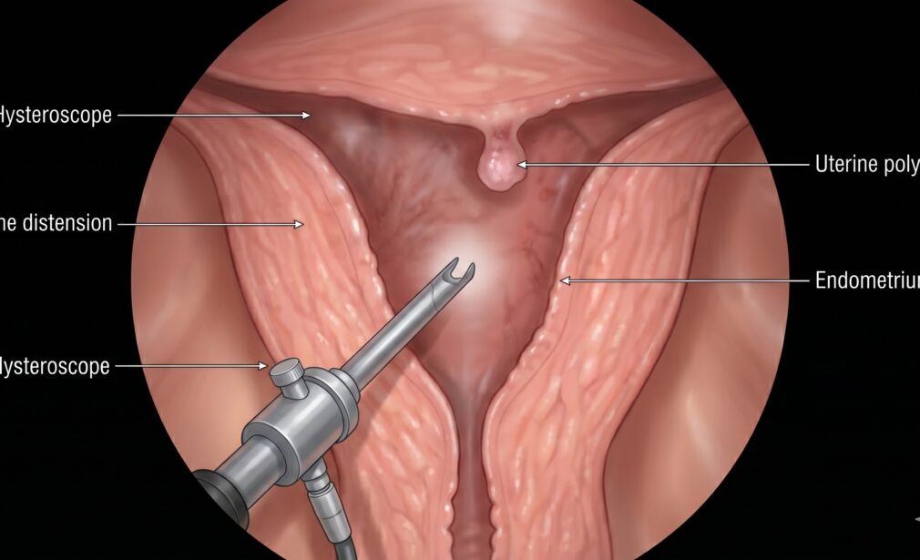
Hysteroscopy
Hysteroscopy is a procedure used to examine the inside of the uterus (the uterine cavity) and the lining of the womb (endometrium). It is unique because it requires no external incisions, utilizing the body’s natural pathways.
Diagnostic vs. Operative Hysteroscopy
Diagnostic: Used to confirm findings from an ultrasound, such as an irregular uterine shape or suspected polyps. It can often be done in an office setting with minimal or no anesthesia.
Operative: Performed in an operating theater under sedation. The surgeon uses a “Resectoscope”—a hysteroscope equipped with a wire loop—to surgically remove abnormalities.
The Procedure
The surgeon dilates the cervix slightly and inserts the hysteroscope through the vagina. A saline solution is pumped into the uterus to expand the walls, providing a panoramic view. This allows for the precise removal of uterine septums (congenital walls inside the uterus) or submucosal fibroids that cause heavy bleeding.
Recovery and Benefits
Recovery is rapid. Most women experience mild cramping (similar to a period) and light spotting for a few days. There is no external scarring, and the risk of infection is lower than with abdominal procedures.